Neurobiology of Sleep-Wakefulness Cycle 1(1): 1 - 8, 2001

DOES HIPPOCAMPAL THETA EXIST IN THE HUMAN BRAIN?

Sunao Uchida, Masaki Nishida, Nobuhide Hirai, Fumikazu Miwakeichi

Department of Sleep Disorders Research, Tokyo Institute of Psychiatry, Tokyo

Accepted in revised form 12 October 2001; recieved 28 August 2001

 
  Summary | Introduction | References | Figures |

Summary

In several species of animal, a regular rhythm known as the hippocampal theta oscillation has been described. However, in humans, the presence of a comparable oscillation has not been established. In animal studies, the phase of theta activity has been related to important roles in the induction of LTP/LTD at the cellular level. The occurrence of theta has also been suggested to reflect the gating of memory related information processing. Thus, the presence or absence of theta oscillation in the human hippocampus has become an essential question. However, past studies have reported contradictory results. Recent results from our laboratory suggest the presence of beta-1 frequency oscillation, rather than theta is present in the human medial temporal lobe. A separate observation indicates that theta oscillation can be observed in the human anterior cingulate cortex. Implications of these findings for conceptualizing human memory functions, and their physiological significance are discussed.

Key Words: Human hippocampus, Electrocorticogram, Beta-1 oscillation, Theta oscillation, Rhythmic slow activity

 
  Summary | Introduction | References | Figures |

Introduction

The ultimate goal of neuroscientific studies is the elucidation of the function and neuronal mechanisms of the human brain. Recent progress in non-invasive methods (e.g. fMRI, PET, MEG, multi-channel EEG) have been very successful in localizing anatomical structures involved during specific brain functions. However, human studies usually pose extreme difficulties for detailed direct examinations of functioning brain tissue. There are still many areas of research (i.e. physiological research) in which the living human brain cannot be directly examined. In such cases, a great deal of knowledge has been obtained from the use of experimental animals.

The greatest advantage of animal studies has come from opportunities to invasively study non-human species. Thus, for example, most electrophysiological studies of neuronal functions have been done using animal subjects. However, a major problem with many animal studies occurs when the animals cannot be shown to behave like humans. Animal models of human psychosis offer an extreme example. Further, there is no guarantee that analogous structures in an animal's brain are homologous with similar structures in the human brain. Thus, both human data and animal data must always be carefully compared and examined in detail to avoid incorrect conclusions about human brain functions.

In physiological studies of the hippocampus, a theta range (4-7 Hz) rhythmic oscillation known as the hippocampal theta oscillation or rhythmic slow activity (RSA) has been observed in many kinds of animals, which has drawn the attention of neurobiologists because of its possible functional role in memory. However, its presence in the human brain has not been established. The hippocampal structures are also frequently the foci of human epileptic activity. Thus, in humans, data from intra-cranial electrodes are occasionally collected from this region for clinical purposes. These cases offer rare opportunities to directly examine the electrophysiology of the human brain.

This article briefly reviews the physiological studies of the hippocampal theta oscillation, and then incorporates our recent data from human temporal and frontal lobe electrophysiological recordings. By comparing human and animal studies; and considering their implications, clues emerge for understanding human hippocampal physiology.

Hippocampal theta oscillation

The hippocampal theta oscillation was first described by Jung and Kornmueller (1938), who found a 5-6 Hz regular rhythmic oscillation in the unanesthetized rabbit hippocampus. Later, Green and Arduini (1954) reported hippocampal theta in both rabbits and cats in the wake state. Both reports indicated that the theta activity was enhanced by painful stimuli. A sleep researcher reported the next notable discovery of the hippocampal theta oscillation. Jouvet et al. (1959) reported that in cats the theta rhythm was a prominent feature of hippocampal activity during paradoxical (rapid eye movement (REM)) sleep. Almost simultaneously, Shimazono (1960) independently reported the same finding in dogs. Theta EEG activity has become one of the criteria for scoring sleep stages in animals. Since the medial temporal lobe was known to play an important role in mnestic functions after the case report of HM (Scoville and Milner 1957), these findings were soon combined to support a possible hippocampal function in memory.

Functional significance of Hippocampal theta oscillation

Independent of the discovery of the theta oscillation, another interesting physiological phenomenon of hippocampal tissue has been described. Bliss and Lomo (1973) reported that in an isolated slice of hippocampal tissue, brief trains of high-frequency stimulation to monosynaptic excitatory connections caused an abrupt and sustained increase in the efficacy of synaptic transmission, reflected concretely by increases in the amplitude of excitatory postsynaptic potentials. The effect occurs within milliseconds of stimulation and can persist for hours and sometimes days. The effect (long-term potentiation (LTP)) has been considered to be a physiological base of memory (see Bliss and Collingridge 1993).

The high frequency stimulation that was typically used to induce LTP was a train of 50-100 stimuli delivered at or above 100 Hz. Interestingly, specially designed short bursts of high frequency (i.e. 4 pulses at 100 Hz) delivered at 200 ms interval (i.e. the theta band) were found to induce LTP more efficiently (Larson et al. 1986). This evidence of the efficacy of 'theta burst stimulation' strongly suggests a physiological relationship between the hippocampal theta oscillation and the induction of LTP.

If theta timing is controlled by the hippocampal theta generation mechanism, how might that high frequency stimulation arise in vivo? Although a physiological equivalent for in vivo high frequency LTP stimulation has not been established, there have been several studies that support its existence (Fox and Ranck 1981; Steward et al. 1992; Bragin et al. 1995). Such findings suggest that the theta oscillation and high frequency activity work together to induce LTP in the hippocampus.

Konopacki et al. (1987) reported the induction of rhythmic theta oscillation in a rat hippocampal slice by soaking it in a carbachol bath. That finding was important because it has enabled researchers to examine both the theta oscillation and LTP induction in vitro. Huerta and Lisman (1993) presented high frequency short burst (4 pulses at 100 Hz) stimuli during various phases of carbachol induced theta waves and found efficiently induced LTP when stimulation occurred during the peak of theta. By contrast, long-term depression (LTD: long-term decreased efficacy of synaptic transmission- see (Kemp and Bashir 2001)) resulted when stimuli were delivered during the trough of theta waves. Poe et al. (2000) examined the relationships between the firing of hippocampal neurons and the phase of the theta rhythm in rats during REM sleep. They found that when rats were in a familiar circumstance, the firing of hippocampal neurons tended to occur in the troughs of theta waves during the following REM sleep, which would thus be likely to induce LTD. However, after rats were exposed to novel circumstances, firing tended to occur during the peaks of theta waves, and thus likely to induce LTP. Poe at al. (2000) concluded that useless memories acquired during familiar circumstance were deleted during subsequent REM sleep, while useful new memories acquired during novel circumstances were strengthened.

O'Keefe had earlier suggested the importance of theta phase, having originally found that the hippocampus is linked to spatial navigation with the discovery of 'place cells' in the hippocampus of freely moving rats (O'Keefe and Dostrovsky 1971). Recently, they observed that when a rat runs through the receptive field of a place cell, the place cell fires bursts of spikes with an inter-burst frequency slightly higher than that of the concomitant theta EEG. This is probably the firing burst is present on slightly earlier phase than preceding theta wave, so that the burst firing point of theta phase may correlate with the animal's location (O'Keefe and Burgess 1999).

Lisman and Idiart (1995) hypothesized that theta phase reflects the activity of a unit of memory processing. Since the gamma frequency is generally considered as about 40 Hz and theta frequency is 5-12 Hz, the maximum number of gamma waves that can occur during a theta wave is approximately seven. Lisman and Idiart (1995) hypothesized that since the number of items a human can store in short term memory is also around seven, the theta oscillation could reflect a timing mechanism for controlling the serial processing of short-term memories.

Such findings strongly suggest that the hippocampal theta oscillation plays an essential role in inducing LTP. The strength of these findings also suggests that if an animal does not exhibit a theta oscillation, there will be no homologous LTP mechanism present in the animal's hippocampus. Thus, establishing the presence or absence of theta oscillation in the human hippocampus has become an essential question.

Human hippocampal oscillation

The hippocampal theta oscillation was originally found in rodents (Jung and Kornmueller 1938). Although they primarily presented the existence of hippocampal theta oscillations in rabbits and cats, Green and Arduini (1954) also examined hippocampal theta oscillations in monkeys, but only briefly described that the "responses in monkey are weak (p. 541)".

Later, several reports on human medial temporal lobe (MTL) electrical activities were reported. It was found that electrically stimulating of the posterior hypothalamus in human produces theta activity in the hippocampus (Sano et al. 1970). Freemon and Walter (1970) examined twelve records from eight patients with depth electrodes, and found that limbic signals, similar to those from neocortex, showed low-voltage fast patterns during REM sleep. Those authors also reported that low-voltage fast activity was observed in some limbic sites during stage 4 sleep. Halgren et al. (1978) found 5-6 Hz slow waves in recordings from one patient's hippocampus. However, that theta activity was reduced by task performance. They concluded that the activity was almost certainly abnormal since it had not been found in other patients. To the contrary, Arnolds et al. (1980) performed spectral analysis using a series of filters (1 Hz width) and found a theta (3-4 Hz) peak; they found that, in one subject, intense behavior enhanced the theta components in hippocampal signals. Using deep electrodes (4 mm above the hippocampal pes), Giaquinto (1973) also reported 5 Hz theta activity during REM sleep, but only in one patient. More recently, Meador et al. (1991) conducted a similar study with a larger sample size. Using recordings from sixteen epileptic patients, they performed power spectral analysis of hippocampal signals from deep electrodes. Power spectra were classified into delta (1-3.75 Hz), theta (4-7.75 Hz), alpha (8-12.75 Hz) and beta (13-30 Hz) frequency bands. During a resting condition while patients' eyes were closed, and then during an auditory word task while patients' eyes were open, theta spectral power was increased compared to resting eyes open and eyes open during visuospatial tasks. Although Meador et al. (1991) concluded that their data were consistent with Arnolds' (1980) results, there was a substantial difference in frequency band definitions between the two studies. Theta peaks in Arnolds' (1980) subjects occurred at 3-4 Hz, while Meador's theta range was 4-7.75 Hz. Since Meador did not present raw data and did not perform narrower band analyses, it remains unclear whether their spectral peak occurred in the same theta range.

Human medial temporal lobe and anterior cingulate cortex activities

Since the early description of the hippocampal theta oscillation, the electrical manifestations of human hippocampal activity has drawn the interest of neuroscientists. One of the earliest papers of animal hippocampal theta activity by Green and Arduini (1954) noted its apparent relation to human EEG: "It is evident that a certain similarity exists between the hippocampal arousal response and the 4-7 / sec theta rhythm described by Walter in the human electrocorticogram of children and psychopaths (p. 553)." Yet, despite the later human studies reviewed above, evidence for the presence of the hippocampal theta oscillation in the human hippocampus has remained contradictory.

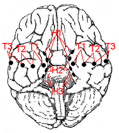
Our group has been studying the electrophysiological properties of the human brain using electrocorticogram (ECoG) recordings of epileptic patients being evaluated for possible neurosurgical treatments. Since most of these patients exhibited medial temporal epilepsy, in the majority of cases electrodes were attached to the medial temporal lobe (MTL). The recording electrode positions are indicated in Figure 1. The electrode assembly was originally developed for recording MTL and basal temporal lobe (BTL) electrocorticograms (EcoG), and has been consistently placed in clinical examinations (Shimizu et al. 1992). Consistent use of the same electrode design and placement for the recording ECoG has provided exceptional opportunities to study the electrophysiological properties of the human MTL.

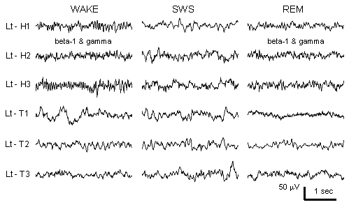
The EcoG data thus obtained indicate that during wakefulness the signals from the human MTL demonstrate two distinct frequency oscillations, beta-1 (10-20 Hz) (Hirai et al. 1999), and gamma (30-150 Hz) (Hirai et al. 1999). Since the hippocampal theta rhythm in non-human animals changes between NREM and REM sleep, we examined how these frequencies change across all-night sleep in humans (Uchida et al. 2001) and found that the beta-1 oscillation was also present during REM sleep (see Figure 2). By contrast, gamma activity was present in all sleep and awake states, although it slightly decreased during slow wave sleep. Thus, we failed to find a 'theta' band oscillation in the human MTL. Nevertheless, activity in the beta-1 band did exhibit characteristics similar to animal hippocampal theta, appearing during both wakefulness and REM sleep. We hypothesized that the human beta-1 may be a functional equivalent of the animal hippocampal theta rhythm.

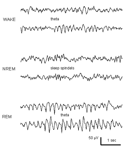
Recently, we studied a patient with electrodes attached to the anterior cingulate cortex (ACC) (Uchida et al. in submission). This was a patient who had previously manifested gamma and beta-1 oscillations in the medial temporal cortex. Thus, we could simultaneously examine both MTL and ACC electrical activities during all-night sleep in one patient. This patient showed gamma and beta-1 oscillations in the MTL, as we previously reported. In the same patients, a separate recording from ACC showed very regular and continuous theta oscillations during wakefulness and REM sleep, but not during NREM sleep (see Figure 3). The ACC FFT power spectra showed corresponding peaks in the theta (5-6 Hz) band during both wake and REM. The waking theta oscillation was independent of whether the eyes were open or closed. A simultaneously recorded Cz EEG showed a regular alpha rhythm during wake, which was suppressed by eyes opening. Therefore, the theta oscillation in the ACC appeared independent of the EEG alpha rhythm.

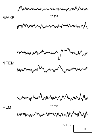
Since we have only been able to study one patient with electrodes attached to both MTL and ACC, we have not yet been able to confirm that ACC theta is commonly observable in humans. However, theta oscillation (Figure 4) has been found in other patients with electrodes placed in the orbitofrontal cortex (OFC). Thus, it seems likely that ACC theta could also be observed in many cases.

Implication of our observation

Functional neuro-imaging studies and lesion studies both suggest that the MTL is important for spatial memory in humans (Maguire et al. 1999). Since it has been suggested that the hippocampal theta oscillation has an essential role in the spatial memory of several species, some equivalent oscillation should be expected in the human MTL. Our observations indicate that the beta-1 (10-20 Hz) frequency in the human MTL may reflect brain processes comparable to those underlying animal theta. If the analogy holds, it suggests that the memory processing cycle occurs substantially faster in the human brain. Since Eichenbaum et al. proposed the hippocampus is not merely processing spatial memory, but forming relational representations between all kinds of stimuli (Eichenbaum et al. 2000; Suzuki and Clayton 2000), the functions of MTL activity may also be related to such functions.

What, then, is the functional role of the ACC theta oscillation we observed? Using cortical electrodes, Kahana et al. (1999) reported clearly enhanced theta oscillation in the raw cortical signals from various parts of the human cerebral cortex during virtual maze navigation in humans. Unfortunately, there were no electrodes attached to the MTL in that study, and it remains unclear whether the theta activity was originated from the MTL. In light of our observations, it seems possible the theta recorded by Kahana et al. (1999) could have originated, perhaps partially, from the ACC. It is also known that a regular frontal midline theta (Fm theta) oscillation is observed in surface EEG recorded during mental concentration (Ishihara and N.Yoshii 1972). That Fm theta could represent the same activity seen as ACC theta in our observation. Reports of non-invasive estimation using EEG or MEG have also suggested the presence of theta band activities in the ACC (Gevins et al. 1997; Ishii et al. 1999), or MTL (Tesche 1997).

Regardless of the specific locus of its origin, it is accepted that the theta oscillation is enhanced by spatial navigation tasks. If theta originates from the ACC, what functional role could the ACC play in spatial memory? Since Fm theta is enhanced by a mental task, and the ACC is known to carry a role in attention or error detection (Bush et al. 2000), ACC theta could be a correlate of functions complementary to spatial (or more extended) memory. Vertes et al. (2001) suggested that the theta rhythmic signal resonates throughout Papez' circuit, which involves the hippocampus, mammillary body, anterior thalamus, cingulate cortex, and parahippocampal gyrus. Thus, the circuit including both MTL and the ACC may be necessary for efficient memory functions.

Epilogue

Although the importance of the hippocampus in memory function is now certain, present knowledge is still far from completely explaining its physiological function in memory. Such functional relationships are further veiled by the physiological differences between the brains of humans and other more readily studied species. In the present commentary, we have attempted to organize present knowledge to elucidate what is now known and to emphasize again the importance of human studies for understanding human brain function. We plan to pursue such questions in future studies, which may contribute to understanding the functional similarities and differences in the brain electrophysiology of humans and animals.

 
  Summary | Introduction | References | Figures |

References

Arnolds, D.E.A.T., Lopes da Silva, F.H., Aitink, J.W., Kamp, A. and Boeijinga, P. The spectral properties of hippocampal EEG related to behaviour in man. Electroenceph. clin. Neurophysiol., 1980, 50: 324-328.

Bliss, T.V.P. and Collingridge, G.L. A synaptic medel of memory: long-term potentiation in the hippocampus. Nature, 1993, 361: 31-39.

Bliss, T.V.P. and Lomo, T. Long-lasting potentiation of synaptic transmission in the dentate area of the anaesthetized rabbit following stimulation of the perforant path. J. Physiol., Lond., 1973, 232: 331-356.

Bragin, A., Jando, G., Nadasdy, Z., Hetke, J., Wise, K. and Buzsaki, G. Gamma (40-100 Hz) oscillation in the hippocampus of the behaving rat. J. Neurosci., 1995, 15: 47-60.

Bush, G., Luu, P. and Posner, M.I. Cognitive and emotional influence in anterior cingulate cortex. Trends in Cognitive Sciences, 2000, 4: 215-222.

Eichenbaum, H. A cortical-hippocampal system for declarative memory. Nature Reviews Neuroscience, 2000, 1: 41-50.

Fox, S.E. and Ranck, J.B. Electrophysiological characteristics of hippocampal complex-spike cells and theta cells. Experimental Brain Research., 1981.

Freemon, F.R. and Walter, R.D. Electrical activity of human limbic system during sleep. Comprehensive Psychiatry, 1970, 11: 544-551.

Gevins, A., Smith, M.E., McEvoy, L. and Yu, D. High-resolution EEG mapping of cortical activation related to working memory: effects of task difficulty, type of processing, and practice. Cereb. Cortex, 1997, 7: 374-385.

Giaquinto, S. Sleep recordings from limbic structure in man. Confin. Neurol., 1973, 35: 285-303.

Green, J.D. and Arduini, A. Hippocampal electrical activity in arousal. J. Neurophysiol., 1954, 17: 533-557.

Halgren, E., Babb, T.L. and Crandall, P.H. Human hippocampal formation EEG desynchronizes during attentiveness and movement. Electroenceph. clin. Neurophysiol., 1978, 44: 778-781.

Hirai, N., Uchida, S., Maehara, T., Okubo, Y. and Shimizu, H. Beta-1 (10-20 Hz) cortical oscillations observed in the human medial temporal lobe. Neuroreport, 1999, 10: 3055-3059.

Hirai, N., Uchida, S., Maehara, T., Okubo, Y. and Shimizu, H. Enhanced gamma (30-150 Hz) frequency in the human medial temporal lobe. Neuroscience, 1999, 90: 1149-1155.

Huerta, P.T. and Lisman, J.E. Heightened synaptic plasticity of hippocampal CA1 neurons during a cholinergically induced rhythmic state. Nature, 1993, 364: 723-725.

Ishihara, T. and N.Yoshii Multivariate analytic study of EEG and mental activity in juvenile delinquents. Electroenceph. clin. Neurophysiol., 1972, 33: 71-80.

Ishii, R., Shinosaki, K., Ukai, S., Inouye, T., Ishihara, T., Yoshimine, T., Hirabuki, N., Asada, H., Kihira, T., Robinson, S.E. and Takeda, M. Medial prefrontal cortex generates frontal midline theta rhythm. Neuroreport, 1999, 10: 675-679.

Jouvet, M., Michel, F. and Courjon, J. Sur un stade dactivite electrique cerebrale rapide au cours du sommeil physiologique. C. R. Soc. Biol., 1959, 153: 1024-1028.

Jung, R. and Kornmuller, A. Ein methodik der abteilung lokalsierter potential schwankingen aus subcorticalen hirnyebieten. Arch. Psychiat. Neuroenkr., 1938, 109: 1-30.

Kahana, M.J., Sekuler, R., Caplan, J.B., Kirschen, M. and Madsen, J.R. Human theta oscillations exhibit task dependence during virtual maze navigation. Nature, 1999, 399: 781-784.

Kemp, N. and Bashir, Z.I. Long-term depression: a cascade of induction and expression mechanisms. Progress in Neurobiology, 2001, 65: 339-365.

Konopacki, J., MacIver, M.B., Bland, B.H. and Roth, S.H. Carbachol-induced EEG "theta" activity in hippocampal brain slices. Brain Res., 1987, 405: 196-198.

Larson, J., Wong, D. and Lynch, G. Patterned stimulation at the theta frequency is optimal for the induction of hippocampal long-term potentiation. Brain Res., 1986, 368: 347-350.

Lisman, J.E. and Idiart, M.A.P. Storage of 7 plus-minus 2 short-term memories in oscillatory subcycles. Science, 1995, 267: 1512-1515.

Maguire, E.A., Burgess, N. and O'Keefe, J. Human spatial navigation: cognitive maps, sexual dimorphism, and neural substrates. Current Opinion in Neurobiology, 1999, 2: 171-177.

Meador, K.J., Thompson, J.L., Loring, D.W., Murro, A.M., King, D.W., Gallagher, B.B., Lee, G.P., Smith, J.R. and Flanigin, H.F. Behavioral state-specific changes in human hippocampal theta activity. Neurology, 1991, 41: 869-872.

O'Keefe, J. and Burgess, N. Theta activity, virtual navigation and the human hippocampus. Trends in Cognitive Science, 1999, 3: 403-406.

O'Keefe, J. and Dostrovsky, J. The hippocampus as a spatial map: preliminary evidence from unit activity in the freely moving rat. Brain Res., 1971, 34: 171-175.

Poe, G.R., Nitz, D.A., McNaughton, B.L. and Barnes, C.A. Experience-dependent phase-reversal of hippocampal neuron firing during REM sleep. Brain Research, 2000, 855: 176-180.

Sano, K., Mayanagi, Y., Sekino, H., Ogashiwa, M. and Ishijima, B. Results of stimulation and destruction of the posterior hypothalamus in man. J Neurosurg, 1970, 33: 689-707.

Scoville, W.B. and Milner, B. Loss of recent memory after bilateral hippocampal lesions. Journal of Neurology, Neurosurgery and Psychiatry, 1957, 20: 11-21.

Shimazono, Y., Horie, T., Yanagisawa, N., Hori, N., Chikazawa, S. and Shozuka, K. The correlation of the rhythmic waves of the hippocampus with the behaviors of dogs. Neurol. med. -chirur., 1960, 2: 82-88.

Shimizu, H., Suzuki, I., Ohta, Y. and Ishijima, B. Mesial temporal subdural electrode as a substitute for depth electrode. Surg. Neurol., 1992, 38: 186-191.

Steward, M., Luo, Y. and Fox, S.E. Effects of atropine on hippocampal theta cells and complex-spike cells. Brain Res., 1992, 591: 122-128.

Suzuki, W.A. and Clayton, N.S. The hippocampus and memory: a comparative and ethological perspective. Current Opinion in Neurobiology, 2000, 10: 768-773.

Tesche, C.D. Non-invasive detection of ongoing neuronal population activity in normal human hippocampus. Brain Res., 1997, 749: 53-60.

Uchida, S., Maehara, T., Hirai, N., Kawai, K. and Shimizu, H. Theta oscillation in the anterior cingulate and beta-1 oscillation in the medial temporal cortices: A human case report. (Submitted).

Uchida, S., Maehara, T., Hirai, N., Okubo, Y. and Shimizu, H. Cortical oscillations in human medial temporal lobe during wakefulness and all-night sleep. Brain Res., 2001, 891: 7-19.

Vertes, R.P., Albo, Z. and Viana di Prisco, G. Theta-rhythmically firing neurons in the anterior thalamus: Implications for mnemonic functions of Papez's circuit. Neuroscience, 2001, 104: 619-625.
 
  Summary | Introduction | References | Figures |

Figures

Figure 1. Positions of electrodes placed for medial and basal temporal lobe recordings.
Montage identifications are also indicated.


Figure 2. Raw signals from medial and basal temporal lobe electrode sites.
Montages are as indicated for Figure 1. The medial temporal lobe signals demonstrate beta-1 and gamma oscillations during wake and REM sleep. High frequency gamma oscillation is superimposed on the signals, most clearly manifested during SWS H2, and H3.


Figure 3. Raw signals from the patient's right anterior cingulate cortex.
A regular theta oscillation is observed during wake and REM sleep, but not during NREM sleep. During NREM sleep, sleep spindles are apparent.


Figure 4. Raw signals from the patient's right orbito-frontal cortex.
Weak, but regular theta oscillation occurred during wake and REM sleep. (Unpublished data).


Correspondence: Uchida Sunao, MD., PhD.,
Department of Sleep Disorders Research,
Tokio Institute of Psychiatri,
2-1-8 Kamikitazawa, Setagaya-ku, Tokio, 156-8585, Japan.
E-mail: sunao@uchidae.com